Chest And Abdominal Muscles Diagram / Rectus Abdominis - Abdominal Muscles - Anatomy Muscles ... : The abdominal wall surrounds the abdominal cavity, which houses several abdominal structure and.. Respiratory muscle training online course: Most will label a diagram of muscle with its structures. The muscles are divided by a ligament running posteriorly from the axis and along the midline the thorax, or chest, is the superior part of the trunk situated between the neck and abdomen. The diaphragm forms the upper surface of. Muscles extend from the iliac crest to inferior border of the ribs, they are positioned 3.
The abdominal wall encloses the abdominal cavity, which holds the bulk of the gastrointestinal viscera. Learn vocabulary, terms and more with flashcards, games and other study tools. Muscles of the abdominal wall. Learn about its function, parts, abdominal conditions, and more. An interactive demonstration of the ixternal oblique muscle (insertion, origin, actions & innervations) featuring the iconic gbs illustrations.
Muscle Chart Male | Body muscle chart, Abdominal muscles ... from i.pinimg.com The anterior muscles of the trunk (torso) are associated with the front of the body, include chest and abdominal muscles. Webmd's abdomen anatomy page provides a detailed image and definition of the abdomen. Groin muscles diagram anterior muscles diagram muscle diagram anterior muscular system. Contraction of the diaphragm causes it to descend towards the abdomen, increasing the space of the thoracic cavity and expanding the lungs, filling them with air. Abdominal exercises can still be beneficial to your program. Two sphincter muscles control the anus; The abdominal wall encloses the abdominal cavity, which holds the bulk of the gastrointestinal viscera. Thoracic wall and abdominal cavity.
Muscles extend from the iliac crest to inferior border of the ribs, they are positioned 3.
Although the abdominal muscles have intersegmental nerve stimulation, you are not able to contract one section independent of the other. Everyone should list the structures within muscle. Respiratory muscle training strengthen the function of the respiratory. Related online courses on physioplus. The abdomen (colloquially called the stomach, belly, tummy or midriff) is the part of the body between the thorax (chest) and pelvis, in humans and in other vertebrates. The abdominal wall encloses the abdominal cavity, which holds the bulk of the gastrointestinal viscera. Together, those muscles act mainly to flex the hip, but they also contribute to abdominal flexion and hip stabilization. It is a broad and thin muscle with its muscular portion covering the side and aponeurosis on the anterior wall. This page provides an overview of the chest muscle group. Learn about its function, parts, abdominal conditions, and more. Fabian identifying the muscles and landmarks of the abdomen and chest. In the hanging leg lift, the rectus abdominis must rotate the pelvis posteriorly and stabilize the pelvis to allow the legs to move freely toward the chest. Start studying chest and abdominal muscles.
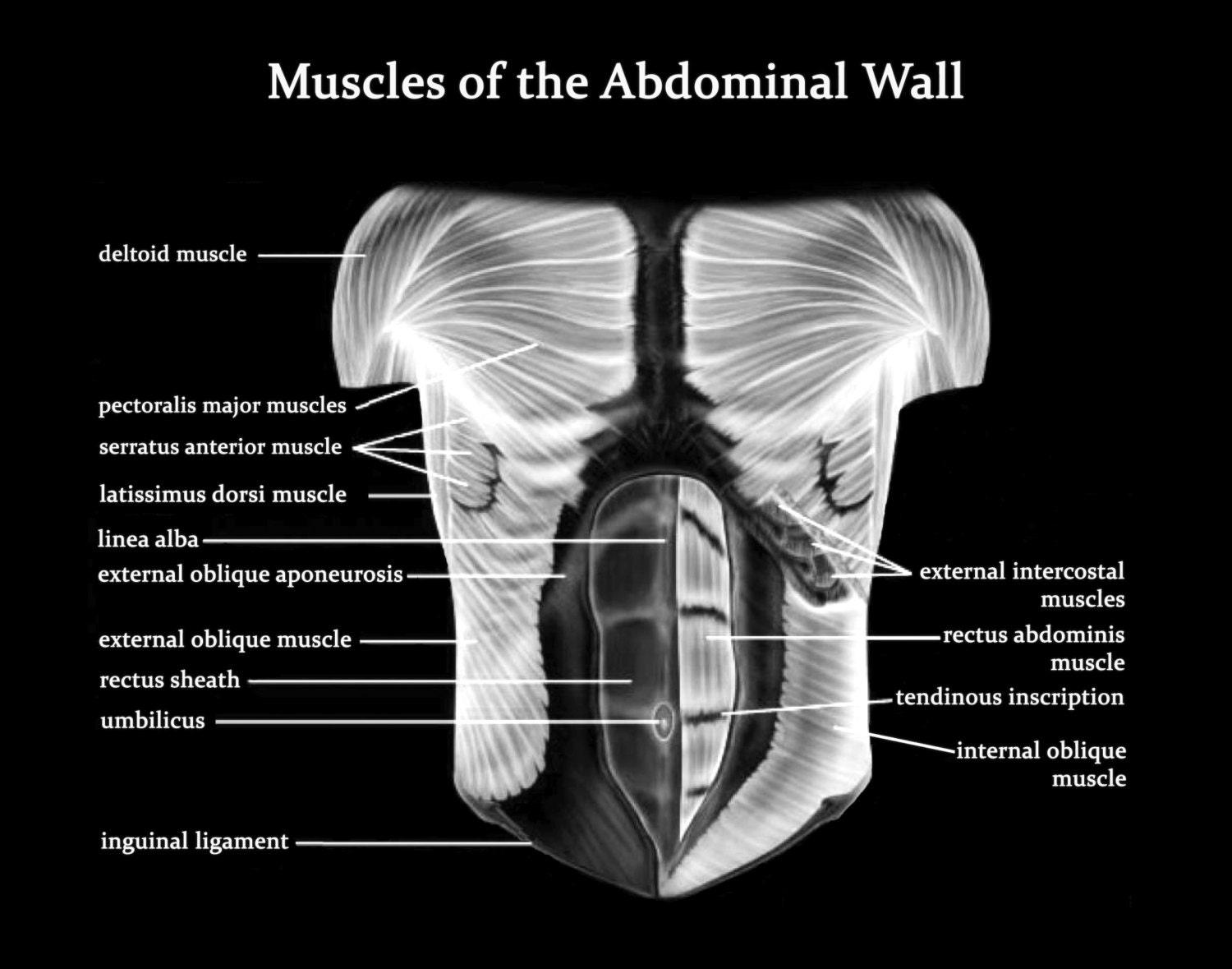
The abdominal wall surrounds the abdominal cavity, which houses several abdominal structure and. The abdominal muscles form the anterior and lateral abdominal wall. Here is a diagram that shows where each one is located: Its main roles are to stabilise the trunk and maintain internal abdominal pressure. Thoracic wall and abdominal cavity.
Muscles of the Abdominal Wall Art Print Poster Medical from img0.etsystatic.com Thoracic wall and abdominal cavity. Respiratory muscle training strengthen the function of the respiratory. Most will label a diagram of muscle with its structures. Two sphincter muscles control the anus; Respiratory muscle training online course: You just want to make sure you choose the most effective moves. It is a broad and thin muscle with its muscular portion covering the side and aponeurosis on the anterior wall. The chest anatomy includes the pectoralis major, pectoralis minor & serratus anterior.
This muscle forms the anterior and lateral abdominal wall.
Respiratory muscle training online course: Although the abdominal muscles have intersegmental nerve stimulation, you are not able to contract one section independent of the other. The two sides connect at the sternum, or breastbone. Its main roles are to stabilise the trunk and maintain internal abdominal pressure. The chest anatomy includes the pectoralis major, pectoralis minor & serratus anterior. Most will label a diagram of muscle with its structures. Check out this library of free labeling diagrams. Thoracic wall and abdominal cavity. This page provides an overview of the chest muscle group. Start studying chest and abdominal muscles. The muscles are divided by a ligament running posteriorly from the axis and along the midline the thorax, or chest, is the superior part of the trunk situated between the neck and abdomen. Contraction of the diaphragm causes it to descend towards the abdomen, increasing the space of the thoracic cavity and expanding the lungs, filling them with air. It's also important that you know the facts regarding the anatomy and.
The 4 distinct muscles that make up your abs. Its main roles are to stabilise the trunk and maintain internal abdominal pressure. Respiratory muscle training online course: Learn vocabulary, terms and more with flashcards, games and other study tools. Fabian identifying the muscles and landmarks of the abdomen and chest.
torso model muscles with labels - Google Search | Muscle ... from i.pinimg.com The 4 distinct muscles that make up your abs. It's also important that you know the facts regarding the anatomy and. The abdomen (commonly called the belly) is the body space between the thorax (chest) and pelvis. The abdominal wall encloses the abdominal cavity, which holds the bulk of the gastrointestinal viscera. Its main roles are to stabilise the trunk and maintain internal abdominal pressure. Muscles extend from the iliac crest to inferior border of the ribs, they are positioned 3. Related online courses on physioplus. The internal sphincter, consisting of smooth muscle fibers, is under the control of the autonomic nervous system it is situated in the upper part of the abdominal cavity occupying the greater part of the right hypochondriac region, part of the epigastric region, and.
The muscles of this region both allow for this range of motion and contract to stabilize this region and prevent any extraneous motion.
Groin muscles diagram anterior muscles diagram muscle diagram anterior muscular system. The transverse abdominal muscle wraps around the torso from front to back and from the ribs to the pelvis. Now that you have a basic understanding of what the abdominal muscles are and how they work, you can design workouts that actually target these muscles. The abdominal wall encloses the abdominal cavity, which holds the bulk of the gastrointestinal viscera. Start studying chest and abdominal muscles. A layer of muscle and fascia which protects and encloses the abdominal cavity, allowing for its compression as well as torso movement. The anterior muscles of the trunk (torso) are associated with the front of the body, include chest and abdominal muscles. The internal sphincter, consisting of smooth muscle fibers, is under the control of the autonomic nervous system it is situated in the upper part of the abdominal cavity occupying the greater part of the right hypochondriac region, part of the epigastric region, and. Anatomy and attachments of sternum. Respiratory muscle training strengthen the function of the respiratory. Its main roles are to stabilise the trunk and maintain internal abdominal pressure. Although the abdominal muscles have intersegmental nerve stimulation, you are not able to contract one section independent of the other. The muscles are divided by a ligament running posteriorly from the axis and along the midline the thorax, or chest, is the superior part of the trunk situated between the neck and abdomen.
The four main abdominal muscle groups that combine to completely cover the internal organs include: chest muscles diagram. A layer of muscle and fascia which protects and encloses the abdominal cavity, allowing for its compression as well as torso movement.


0 Comments:
Posting Komentar